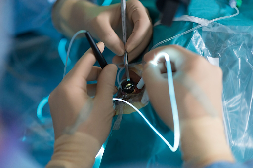
Retinal Detachment Surgery
Retinal detachment surgery is a type of surgical procedure which is performed to repair the condition of retinal detachment. Retinal detachment is a condition in which the retina gets detached from its normal position.
Today, we at MyMedTrip.com shall brief you on retinal detachment surgery, retinal detachment surgery cost in India, and retinal detachment surgery success rate. We will also give you some information on retinal detachment surgery recovery and procedure.
| Particulars | Details |
|---|---|
| Retinal detachment surgery cost in India | 672 USD – 941 USD |
| Discount | 10% on the above quoted price (final hospital’s bill) ONLY APPLICABLE ONLY FOR MyMedTrip.com patients Click here for exceptions and terms. |
| Number of days at hospital (Estimated) | 1 day |
| Number of days in India outside hospital (Estimated) | 15 days |
| Treatment’s Success Rate | 85% |
| Tests required to help assess the treatment | Retinal examination & ultrasound |
What is covered in the above mentioned cost for surgery?
This price includes surgery cost, doctor’s fee, standard prescribed tests and all standard expenses required at the hospital.


Retinal Detachment Surgery

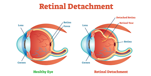
Retina is a thin layer of tissue responsible for sensing light and sending the images to the brain. When retinal detachment occurs, the retinal cells get pulled away from the blood vessels of the eye which are responsible for carrying oxygen.
This condition is symptomized by blurred vision which increasingly becomes prominent. If not treated immediately, retinal detachment can lead to permanent loss of vision. Retinal detachment surgery is a correctional method which is performed to avoid such dire consequences.
In order to understand how retinal detachment occurs and what are its symptoms, let us first try to understand the anatomical structure of the eye. In this train, we will also try to understand the functions of the eye and the causes behind this eye condition, the retinal detachment.
The eyes
The human eye comprises choroid, ciliary body, cornea, fovia, iris, lens, macula,optic nerve, pupil, sclera, vitreous humor, and the retina. Each of these components form the circular structure called the eye.
The cornea lies at the outermost layer of the eyeball’s surface. The ciliary body lies at the corner, right behind the iris. The iris is the round coloured part of our eyeballs responsible for regulating the amount of light that enters the eye. Located within the iris is the pupil.

Behind the pupil and the iris, lies the lenses which directs the light received by the iris, into the retina. With increasing age, these lenses get damaged. When this occurs, special devices such as spectacles or artificial lenses are used to enhance vision.
Connecting the lens to the retina is vitreous humour. The retina is located at the back of the eye and is responsible for transmitting the vision-images into the brains through the optic nerve.
Functions of the eye
The human eye is responsible for transmitting the images we perceive as information, to the brain. It regulates one of the five senses in our body that help us interpret things and understand the world around us.
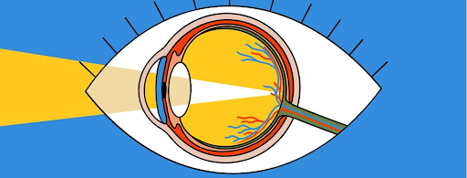
When we see something, our eyes take in the light. The amount of light to be taken in is regulated by the iris. The iris and the lens directs this light to the back of the eye, into the retina. The retina, with the help of the optic nerve, transmits this imagery information to the brain.
Retinal detachment causes and types
There are three types of retinal detachment, rhegmatogenous retinal detachment, tractional retinal detachment, and exudative retinal detachment. Each of these types of retinal detachment are caused by various factors, some of which are noted below.
1. Rhegmatogenous retinal detachment
Rhegmatogenous retinal detachment is the most common type of retinal detachment and occurs with advancing age.
This occurs when a tear in the retina results in the passage of fluid from the inner areas. This fluid is then collected at the back of the retina which then leads to the retina’s separation from the front portion of the eye.
2. Tractional retinal detachment
Tractional retinal detachment, is yet another type of retinal detachment in which the retina detaches itself as a result of scar tissues that grow on its surface.This type is most common among people who have some underlying condition such as diabetes or obesity.
3. Exudative retinal detachment
In the third type of retinal detachment, fluid is accumulated in behind the retina. The retina however does not tear. This condition is called exudative retinal detachment. Exudative retinal detachment occurs when the eye suffers from an injury. A tumour in the eye may also cause this type of retinal detachment.
Symptoms of retinal detachment

The symptoms of retinal detachment are as follows:-
- Blurred vision.
- Appearance of a thinly-veiled curtain, while seeing .
- Reduced peripheral sight.
- Photopsia or light-flashes.
Diagnosis of retinal detachment
Retinal detachment is a serious condition. One must consult the doctor immediately if and when any of these above mentioned symptoms appear. Upon consulting the doctor, the doctor will inquire after the symptoms experienced by the patient.
Tests to be done
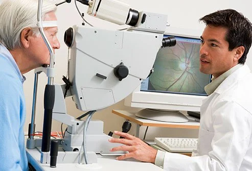
Along with this, the doctor will also suggest some tests such as a retinal examination and an ultrasound imaging of the eye, that need to be done. In a retinal examination, the doctor, with the help of very bright light, examines the internal structures of the eye. This also includes examining the retina.
When the pulling away of the retina creates damage and leads to bleeding, the retinal examination fails to depict the retina clearly. Therefore, the doctor following this, would most likely recommend an imaging test such as the ultrasound imaging test.
Types of retinal detachment surgery
After a retinal detachment has been diagnosed, the doctor will suggest surgical intervention. Surgery is the most effective, most common, and the only way to treat a retinal tear.
Laser surgery and cryopexy
There are various types of retinal detachment surgery. When the retina has not completely detached itself, the ophthalmologist may either recommend a laser surgery or cryopexy.
In a laser surgery, the surgeon, with the help of laser light, destroys the surrounding areas of the retinal tear. In a cryopexy, the surgeon, after numbing the eye with anesthesia, freezes the affected area.
In both the cases, the burning and the freezing, scarring of the retinal area causes the retina to get attached to the eye. Both these kinds of eye surgery are done on an outpatient basis.
Pneumatic retinopexy
This surgery is suggested when the retina has completely detached itself.
In this surgery, the doctor prescribes relaxants which helps the patient relax during the surgery. Following this, the doctor progresses to apply eye drops that will numb the area of the eye and dilate it.
If the doctor feels an additional need to use anesthesia, the doctor will do so by injecting it near the eye. The type of anesthesia will be regional and will only work to numb one specific area.

In the next step, the doctor, with the help of a syringe, will remove some fluid from the area and insert a gas bubble. With the help of special surgical instruments and devices, the doctor will make sure that the gas bubble is right near the retina.
In the following step, the doctor may perform an additional cryopexy to freeze the area and thereby seal the retina in the right location.
After the cryopexy, the doctor completes the surgery by applying antibiotics and sealing up the wounds with the help of bandages.
Scleral buckling
A scleral buckling is suggested when complete retinal detachment has occurred.

The surgery commences under general anesthesia. In some cases, the doctor may also use a regional anesthesia which will numb only one specific area of the body. In this case, the eye.
In the next step, the doctor progresses to apply eye drops which dilate the eyes and makes its structures more visible. Following this, the surgeon progresses to make an incision. The incision is made on the outer layer of the eye.
With the help of an ophthalmoscope, the surgeon is able to view the internal structures of the retina. Through this view, the surgeon progresses to place a small instrument which seals the retina back to its original location.
While placing the ‘freezing’ device, the surgeon drains any excessive fluid that has accumulated behind the detached retina.
The surgeon completes the surgery by applying an antibiotic and sealing up the eye with bandages. An eye patch may also be attached.
Retinal detachment surgery recovery
1. Pneumatic retinopexy
After a pneumatic retinopexy, the patient may wake with some amount of soreness and discomfort. The patient will be provided with an eye patch to avoid any added discomfort or chances of infection. Along with this, the patient will be prescribed antibiotics and pain medications to manage any pain and heal the eye faster.
This retinal detachment surgery recovery comes with a set of instructions in which there is specific information with regards to how to sleep at night. While recovering, the patient will be asked to avoid flights for a few days.
2. Scleral buckling
After a scleral buckling, as in pneumatic retinopexy, the patient may wake with some amount of soreness and discomfort. The patient will be provided with an eye patch to avoid any added discomfort. Along with this, the patient will be prescribed antibiotics and pain medications to manage any pain and heal the eye faster.
Retinal detachment surgery complications
The complications associated with either of the retinal detachment surgeries are:-
- For pneumatic retinopexy- there are complications such as recurrent retinal detachment, gas trapped in the eye, eye inflammation, excessive bleeding in the eye, and new retinal tear.
- For scleral buckling- The risk may be cataracts, double vision, infection, bleeding, new retinal tear, and retinal incarceration.
For a comprehensive list, please ask the ophthalmologist for specific surgery risks.
How can MyMedTrip.com help?
If you have decided to travel to India for retinal detachment surgery, you may contact us on our Whatsapp number +91 9818237391 or email us at hi@mymedtrip.com The first consultation arranged by us is free of cost! We also provide visa invitation letters and help in facilitating the medical journey to India.
Throughout the journey, you shall be provided with one of our staff members for proper guidance through linguistic barriers, even though most of the hospitals and doctors we feature are well versed with Arabic, Russian,Bengali, and English.
If you have any further queries or questions related to retinal detachment surgery services in India, please do not hesitate to email us at the aforementioned address.
Frequently Asked Questions about Retinal Detachment Surgery
What is retinal detachment?
Retinal detachment is a condition in which the retina gets detached from its normal position.
Where is the retina located?
The retina is located at the back of the eye.
What is the function of the retina?
The retina functions to transmit the vision-images into the brains through the optic nerve.
How does retinal detachment occur?
Retinal detachment is a serious condition. It occurs when the retina detaches itself from the wall. As a result of this, the retinal cells get pulled away from the blood vessels of the eye, which are responsible for carrying oxygen.
What are the various types of retinal detachment?
The various types of retinal detachment are rhegmatogenous retinal detachment, tractional retinal detachment, and exudative retinal detachment.
What is the treatment for retinal detachment?
Retinal detachment surgery is the one treatment for retinal detachment.
What tests are required to diagnose retinal detachment?
The tests required to diagnose retinal detachment are a retinal examination and an ultrasound imaging test.
What are the various types of surgeries for retinal detachment?
The various types of retinal detachment surgery are laser surgery, cryopexy, pneumatic retinopexy, and scleral buckling surgery.
What is the procedure for a laser surgery?
In a laser surgery, the surgeon, with the help of laser light, destroys the surrounding areas of the retinal tear
What is the procedure for a cryopexy surgery?
In a cryopexy, the surgeon, after numbing the eye with anesthesia, freezes the affected area.
What happens in a pneumatic retinopexy surgery?
In a pneumatic retinopexy, the surgeon uses a gas bubble to attach the retina to its place
Under what kind of anesthesia does a pneumatic retinopexy take place?
Regional anesthesia.
What happens in a scleral buckling surgery?
In a scleral buckling surgery, the surgeon, through an incision, places a small instrument which seals the retina back to its original location.
What type of anesthesia is used in a scleral buckling surgery?
General anesthesia.