About the Doctor
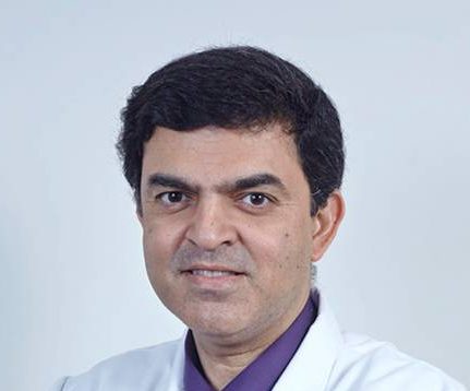
With over 20 years of elaborate experience, Dr. Rajnish Monga specializes in acute pancreatitis and associated gastroenterological disorders. He also specializes in pancreatic and colonic diseases. He has multiple national and international publications in journals with high impact factors. He has expertise in endoscopic procedures including ERCP and EUS.
Specialization
Frequently Asked Questions About Gastroenterology
What is GI Endoscopy?
Gastrointestinal endoscopy is done when to see the inside of the stomach or intestines. Gastrointestinal endoscopy involves placing a thin tube-like instrument through the mouth or the back passage, the rectum, and down into the gullet, or oesophagus, stomach or intestines. The tube is able to carry pictures back to a video screen or camera. When endoscopy is done through the mouth, it is called upper endoscopy or esophagogastroduodenoscopy (EGD for short). When endoscopy is done through the rectum, it is called lower endoscopy or colonoscopy.
Common Reasons For Its Use And Benefits:
- BLEEDING – The most common reason for gastrointestinal endoscopy is to find the source of bleeding from the oesophagus, stomach or intestines. Sometimes, if the source is found, doctors can use the endoscope to stop the bleeding. ?TUMOURS – Sometimes tumours can cause discomfort or even clog the oesophagus, stomach or intestines. The endoscope can be used to find tumours and take a small sample for analysis in the lab, called a biopsy.
- DIARRHEA – Sometimes severe diarrhea can be caused by inflammation or infections of the colon and endoscopy can be used to help find the cause. ?BELLY PAINS – Sometimes severe stomach pain can be a sign of ulcer, a clog in the gastrointestinal track, inflammation or infection. Endoscopy can help find the reasons for some belly pains.
Some of the risks though rare of gastrointestinal endoscopy include:
- A low blood pressure (called hypotension) – Frequently, medicines are given to help keep patients comfortable during endoscopy. Some patients develop brief drops in the blood pressure during endoscopy. Such drops can be life-threatening and can be a reason for stopping endoscopy before it is finished.
- oesophagus, stomach or intestines can be damaged by the endoscope and the contents can leak into the surrounding area of the body. This is a serious problem which can cause infection and even death.
- Bleeding – The oesophagus, stomach or intestines can be damaged by the endoscope causing some internal bleeding.
What is Feeding Tube?
A feeding tube is a medical device used to provide nutrition to patients who cannot obtain nutrition by swallowing. The state of being fed by a feeding tube is called gavage, enteral feeding or tube feeding. Placement may be temporary for the treatment of acute conditions or lifelong in the case of chronic disabilities.
Types Of Feeding Tubes:
- NASOGASTRIC TUBE: NG-tube is passed through the nostril down the oesophagus and into the stomach. This type of feeding tube is generally used for short term feeding, usually only 2 weeks maximum.
- GASTRIC FEEDING TUBE: A gastric feeding tube (G-tube or “button”) is a tube inserted through a small incision in the abdomen into the stomach and is used for long-term enteral nutrition. One type is the Percutaneous endoscopic gastrostomy (PEG) tube which is placed endoscopically. The position of the endoscope can be visualized on the outside of the patient’s abdomen because it contains a powerful light source. These tubes are suitable for long-term use, though they sometimes need to be replaced if used long term. The G-tube can be useful where there is difficulty with swallowing because of neurologic or anatomic disorders and to avoid the risk of aspiration.
- JEJUNOSTOMY FEEDING TUBE: or J-tube is a tube surgically inserted through the abdomen and into the jejunum i.e. the second part of the small intestine.
Complications Of Feeding Tubes
Gastric feeding tubes have a variety of complications that can occur, though the overall rate of complication is about 1%. As gastric feeding tubes are placed as part of a procedure that punches a hole in the stomach and skin, this can lead to leaking of contents into the abdomen. The most frequent complication is irritation around the site of the insertion, generally caused by stomach acid and feedings leaking around the site. Barrier creams, dressings, and frequent cleaning is generally recommended. Especially in advanced dementia, patients can pull at the feeding tubes causing them to be dislodged and requiring a hospitalization to replace them. Feeding tubes may become clogged or occluded if not flushed with water after each feeding. A clogged tube may need to be replaced. Nasogastric feeding tubes, if inserted incorrectly, can cause collapsed lungs.
Withdrawal Of Tubes
Tube feeding, like all medical treatments, can be declined or stopped, especially in the setting of a terminal illness where its use would not alter the ultimate outcome. Alternatively, nutrition can be withheld and the tube used only for hydration and medicine if desired. Some patients or families will opt for a “time limited trial” of feeding through a tube, but after a set time period if the individual is not improving feedings are stopped and the goals of care are refocused to comfort measures
Stomach Tubes
Many critically ill patients are not able to swallow properly. Also, patients on mechanical ventilators cannot eat by mouth. When the stomach and intestines continue to work, a tube is placed through the nose or mouth and pushed down into the stomach. This tube allows nurses to make sure that the stomach does not get over filled, and also to feed the patient. Nasogastric (or “N.G.”) tubes are thicker tubes (about the thickness of a pencil). These tubes are used when it is important both to suck out stomach fluid for testing, to prevent over filling, and for feeding. Feeding tubes are thinner tubes that are used mainly for feeding.
Common Reasons For Its Use And Benefits:
- MONITORING THE STOMACH – This is very important to prevent the stomach from being overfilled with food or stomach juice, and to make sure the stomach juice does not become too acid.
- FEEDING – Some patients who cannot swallow and some patients who are on mechanical ventilators can be fed through nasogastric or feeding tubes.
Risks:
Some of the risks of putting in a nasogastric or feeding tube include:
- DISCOMFORT DURING PLACEMENT – Discomfort can result when the tube is inserted. Doctors try to lessen the pain by putting a jelly on the tube that helps it to slide in more smoothly.
- While the tube is being passed, it can go down the windpipe instead of into the stomach. This can cause coughing. Doctors often get an x-ray to see where the tube goes before they give food or water through it.
- COLLAPSED LUNG – While the tube is being passed, it may, very rarely, go down into the wind pipe and puncture the lung. This hole may seal quickly on its own. If the hole does not seal over, air can build around the lung and cause it to collapse (this is called pneumothorax). In such cases, a chest tube is sometimes needed to drain air from around the lung.
Because of the low risk and common need for stomach tubes, the consent that patients sign for general treatments at the time of coming into the hospital usually includes permission for passing a stomach tube through the nose or mouth if it is needed. If the tube is needed for a long time, doctors may need to make a hole in the abdomen and pass a tube through the skin, into the stomach or intestines. Surgery of this nature requires consent from patients or families.
What is Banding Therapy?
Banding is used to treat oesophageal varices which result from a condition called portal hypertension.
What Is Portal Hypertension ?
Sometimes with liver disease, blood flow through the liver is restricted causing an increase in the blood pressure in and around the liver. This is called portal hypertension. This back pressure of blood can cause a network of enlarged, weak varicose veins to develop in the oesophagus or stomach. These are called varices. If one of these veins ruptures severe bleeding can occur resulting in vomiting of blood or passing blood in the form of black stools. In patients who have fluid retention due to their liver disease, portal hypertension can result in a build up of fluid in the abdomen. This is called ascites.
How Does Banding Work?
Banding is just one way to treat oesophageal varices. It involves a long, flexible tube with a light on the end (an endoscope) which is passed through your mouth into the stomach. The doctor can view the varices in the oesophagus and inserts a rubber band round each of these varices. This stops the blood supply in these veins and they eventually disappear. This will not affect the normal blood supply to the oesophagus. The varices are extra veins that have developed and we need to try to eradicate them, otherwise they may rupture and bleed.
Prior To The Procedure
Banding is sometimes carried out as an ’emergency’ to stop severe bleeding of varices .However sometimes the procedure can be planned ahead of time to prevent the varices from bleeding. In this case, you will be admitted to a ward where blood samples will be taken. The procedure will be explained to you along with the potential risks and you will be asked to sign a consent form. To allow a clear view, the stomach must be empty. You will therefore be asked not to have anything to eat or drink for at least 6 hours before the test. On the day of the banding you will be asked to change into a hospital gown. You will also have to remove any false teeth. A needle will be inserted into a vein in your arm. This is used for giving you sedation during the procedure.
During The Test
- Banding is usually carried out in the endoscopy unit and can take between 15-30 minutes. You will be asked to lie on your left side and will be attached to monitors to measure your heart rate and blood pressure.
- You may be given sedation through the needle in your arm which will make you drowsy and relaxed. The back of your throat will be sprayed to numb the area. To keep your mouth slightly open, a plastic mouthpiece will be gently placed between your teeth.
- The endoscope is then passed into the stomach. This can be a little uncomfortable but is not painful and does not interfere with your breathing. Air is passed down the tube which distends the stomach to allow the doctor to have a better view of the gullet. If you get a lot of saliva in your mouth, this will be cleared using special suction equipment. The bands are then placed around the varices. The air is then sucked out of the stomach and the tube removed quickly and easily.
- Once the procedure is complete, the monitors are removed and you will be transferred back to the ward.
After The Procedure
Once back in the ward you should remain on bed rest until the sedation (if given) has worn off. During this time, nurses will monitor your blood pressure and pulse. You will be able to eat and drink once you are fully awake. However if you have had your throat sprayed, you cannot eat or drink until your swallowing reflex has returned – which takes between 1-2 hours. You may have a sore throat for about 24 hours following the banding, You may also feel a little bloated with the air remaining in your stomach. These discomforts will pass. If the pain is severe or persistent, you should inform the nursing staff.
Are There Any Possible Complications ?
Banding is a safe procedure with a very low frequency of complications, but occasionally the following can occur :
- The banded areas can become scarred, causing a narrowing in your oesophagus and discomfort when you swallow. At worst, this sensation can remain for several weeks, but will resolve.
- Occasionally small ulcers can form in the oesophagus where the banding has been carried out. These can be easily treated with prescribed medication.
- In a very few, rare cases the oesophagus can perforate during the procedure. This may require an operation to reverse.
Follow Up Appointments
- To completely eradicate the varices, you may require several treatments of banding (usually between 3-4), which will require admission to hospital. You will usually be allowed to go home the same day, although occasionally you may need to stay in hospital overnight.
- Once the varices have been successfully treated, you will have an endoscopy every 6 months to 1 year. This is to check the varices have not returned. If varices have developed, they will be banded again.
- It is important that you attend the banding and outpatients appointments. If the varices are left untreated, they may burst and bleed. If you have any signs of bleeding (such as dark stools or vomiting blood) you must inform your doctor immediately.
What is Colonoscopy?
Colonoscopy is a procedure that enables your doctor to examine the lining of the colon (large bowel) by inserting a flexible tube that is about the thickness of your finger into the anus and advancing it slowly into the rectum and colon.Colonoscopy is a procedure that enables your doctor to examine the lining of the colon (large bowel) by inserting a flexible tube that is about the thickness of your finger into the anus and advancing it slowly into the rectum and colon.
What Preparation Is Required?
The colon must be completely clean for the procedure to be accurate and complete. The hospital will give you detailed instructions regarding the dietary restrictions to be followed and the cleansing routine to be used. Preparation consists of either drinking a large volume of a special cleansing solution or several days of clear liquids, laxatives, and enemas prior to the examination. Follow your doctor’s instructions carefully. If you do not, the procedure may have to be cancelled and repeated later.
What About My Current Medications?
Most medications may be continued as usual, but some can interfere with the preparation or the examination. Please inform your doctor of your current medications as well as any allergies to medications several days prior to the examination. Aspirin products, anticoagulants (blood thinners), insulin, and iron products are examples whose use should be discussed with your doctor prior to the examination. You should alert your doctor if you require antibiotics prior to undergoing dental procedures, since you may need antibiotics prior to colonoscopy as well.
What Can Be Expected During Colonoscopy?
Colonoscopy is usually well tolerated and rarely causes much pain. There is often a feeling of pressure, bloating, or cramping at times during the procedure. Your doctor will give you medication through a vein to help you relax and better tolerate any discomfort. The colonoscope is advanced slowly through the large intestine and the lining is carefully examined. The procedure usually takes 15 to 60 minutes. In some cases, passage of the colonoscope through the entire colon cannot be achieved. The doctor will decide if the limited examination is sufficient or if other examinations are necessary.
What If The Colonoscopy Shows Something Abnormal?
If your doctor thinks an area of the bowel needs to be evaluated in greater detail, a forceps instrument is passed through the colonoscope to obtain a biopsy (a sample of the colon lining). If colonoscopy is being performed to identify sites of bleeding, the areas of bleeding may be controlled through the colonoscope by injecting certain medications or by coagulation (sealing off bleeding vessels with heat treatment). If polyps are found, they are generally removed. None of these additional procedures typically produce pain. Remember, the biopsies are taken for many reasons and do not necessarily mean that cancer is suspected.
What Are Polyps And Why Are They Removed?
Polyps are abnormal growths from the lining of the colon which vary in size from a tiny dot to several inches. The majority of polyps are benign (noncancerous) but the doctor cannot always tell a benign from a malignant (cancerous) polyp by its outer appearance alone. For this reason, removed polyps are sent for tissue analysis. Removal of colon polyps is an important means of preventing colorectal cancer.
How Are Polyps Removed?
Tiny polyps may be totally destroyed by fulguration (burning), but larger polyps are removed by a technique called snare polypectomy. The doctor passes a wire loop (snare) through the colonoscope and severs the attachment of the polyp from the intestinal wall by means of an electrical current. You should feel no pain during the polypectomy. There is a small risk that removing a polyp will cause bleeding or result in a burn to the wall of the colon, which could require emergency surgery.
What Happens After A Colonoscopy?
After colonoscopy, your doctor will explain the results to you. If you have been given medications during the procedure, someone must accompany you home from the procedure because of the sedation used during the examination. Even if you feel alert after the procedure, your judgment and reflexes may be impaired by the sedation for the rest of the day, making it unsafe for you to drive or operate any machinery.
What Are The Possible Complications Of Colonoscopy?
- Colonoscopy and polypectomy are generally safe. One possible complication is a perforation or tear through the bowel wall that could require surgery, although this is very uncommon.
- Bleeding may occur from the site of biopsy or polypectomy. It is usually minor and stops on its own orcan be controlled through the colonoscope. Rarely, blood transfusions or surgery may be required.
- Other potential risks include a reaction to the sedatives used and complications from heart or lung disease.
- Localised irritation of the vein where medications were injected may rarely cause a tender lump lasting for several weeks, but this will go away eventually. Applying hot packs or hot moist towels may help relieve discomfort.
What is ERCP?
- It is necessary to have a completely empty stomach. You should therefore fast for at least 6 hours before the procedure.
- If you are allergic to iodine- containing drugs (contrast material or “dye”) you should discuss this with your doctor prior to the procedure.
- The doctor performing the procedure should be informed of any medications that you take regularly, any heart or lung conditions (or any other major diseases), and whether you are allergic to any medications.
- Someone must accompany you home from the procedure because of the sedation used during the examination. Even if you feel alert after the procedure, your judgement and reflexes may be impaired by the sedation for the rest of the day, making it unsafe for you to drive or operate any machinery. If a complication occurs, you may need to stay in hospital until it resolves.
What Can Be Expected During ERCP?
An intravenous sedative will be given to make you more comfortable during the test. Some patients also receive antibiotics before the procedure. The endoscope is passed through the mouth, oesophagus, and stomach into the duodenum. The instrument does not interfere with breathing.
What Are Possible Complications Of ERCP?
- Localised irritation of the vein into which medications were given may rarely cause a tender lump that may last several days.
- Major complications requiring hospitalisation can occur but are uncommon during diagnostic ERCP. They include serious inflammation of the pancreas (‘pancreatitis’) and even more rarely infections, bowel perforation, and bleeding. Another potential risk of ERCP is an adverse reaction to the sedative used. The risks of the procedure vary with the reasons for the test, what is found during the procedure, whether any therapy is undertaken, and the presence of other major medical problems, e.g., heart or lung diseases. Your doctor will tell you what is your likelihood of complications before undergoing the test.
- If therapeutic ERCP is performed (cutting an opening in the bile duct, stone removal, dilation of a stricture (narrowing), stent or drain placement, etc), the possibility of complications is higher than with diagnostic ERCP; complications include pancreatitis, bleeding, and bowel perforation. These risks must be balanced against the potential benefits of the procedure and the risks of alternative surgical treatment of the condition. Often these complications can be managed without surgery, but occasionally they do require corrective surgery.
What Can Be Expected Following ERCP?
If you are having ERCP as an outpatient, you will be kept under observation until most of the effects of the medications have worn off. Evidence of any complications of the procedure will be looked for and hospitalization may be advised if further observation is necessary. You may experience bloating or pass gas because of the air introduced during the examination. You may resume your usual diet unless you are instructed otherwise.
What Is Flexible Sigmoidoscopy?What Is Oesophageal Dilatation?
What Is TIPSS?
What is Flexible Sigmoidoscopy?
Lower Gi Endoscopy Flexible Sigmoidoscopy
Flexible sigmoidoscopy is a procedure that enables your doctor to examine the lining of the rectum and a portion of the colon (large bowel) by inserting a flexible tube that is about the thickness of your finger into the anus and advancing it slowly into the rectum and lower part of the colon.
What Preparation Is Required?
The rectum and lower colon must be completely empty of waste material for the procedure to be accurate and complete. In general, preparation consists of one or two enemas prior to the procedure but may include laxatives or dietary modifications.
What About My Current Medications?
- Most medications can be continued as usual. You should inform your doctor of all current medications as well as any allergies to medications several days prior to the examination.
- However, drugs such as aspirin or anticoagulants (blood thinners) are examples of medications whose use should be discussed with your doctor.
What Can Be Expected During Flexible Sigmoidoscopy?
Flexible sigmoidoscopy is usually well tolerated and rarely causes much pain. There is often a feeling of pressure, bloating, or cramping at various times during the procedure. You will be lying on your side while the sigmoidoscope is advanced through the rectum and colon. As the instrument is withdrawn, the lining of the intestine is carefully examined. The procedure usually takes anywhere from 5 to 15 minutes.
What If The Flexible Sigmoidoscopy Shows Something Abnormal?
If the doctor sees an area that needs evaluation in greater detail, a biopsy (sample of the colon lining) may be obtained and submitted to a laboratory for greater analysis. If polyps (growths from the lining of the colon which vary in size) are found, they can be biopsied, but usually are not removed at the time of the sigmoidoscopy. Polyps are of varying types; certain benign polyps, known as “adenomas,” are potentially precancerous. Certain other polyps (“hyper plastic” by biopsy analysis) may not require removal. Your doctor will likely request that you have a colonoscopy (a complete examination of the colon) to remove any large polyp that is found, or any small polyp that is adenomatous after biopsy analysis.
What Happens After A Flexible Sigmoidoscopy?
- After sigmoidoscopy, the doctor will explain the results to you.
- You may have some mild cramping or bloating sensation because of the air that has been passed into the colon during the examination. This will disappear quickly with the passage of gas.
- You should be able to eat and resume your normal activities after leaving the hospital.
What Are Possible Complications Of Flexible Sigmoidoscopy?
Possible compl icat ions af ter f lexible sigmoidoscopy are rare but few complications may occur—
- Severe abdominal pain,
- Fevers and chills, or
- Rectal bleeding .It is important to note that rectal bleeding can occur even several days after the biopsy.
What is Oesophageal Dilatation?
The oesophagus is a muscular tube that pushes the food from the mouth to the stomach. If the gullet gets narrowed, swallowing becomes difficult and food intake can be severely impaired. Then the narrow part has to be stretched up to allow proper swallowing. The procedure to stretch the gullet is called oesophageal dilatation.
Who Needs An Oesophageal Dilatation?
Usually prior to stretching of your gullet other tests such as a diagnostic endoscopy (camera test) or a barium swallow (x-ray of the gullet) have shown that your gullet has become narrowed. Usually this is the result of severe acid reflux from the stomach into the gullet causing acid burn and scarring, although in some cases it can be due to a growth in the gullet or the result of previous surgery in the oesophagus. Such individuals could be advised oesophageal dilatation.
Who Will Be Doing The Procedure And Where?
A specialist Gastroenterologist with expertise in the procedure will be doing your test. The procedure is usually done in the Endoscopy Unit like any other camera test.
What Is The Preparation For The Procedure?
- You need to be in hospital a few hours prior to the procedure to have a routine clinical examination and some blood investigations done.
- You will be asked to fast for four to six hours prior to the procedure.
- A sedative and a painkiller will be given intravenously just before the procedure to ensure that you are kept comfortable throughout the test.
- You will be asked to put on a hospital gown and sign the concent form.
- You should continue all your medications but if you take any medications that make your blood thinner (anticoagulants) such as warfarin, or if you’re diabetic on insulin you must let your doctor know at least 3 days in advance.
- If you have any allergies you must let the nursing staff and doctors know.
What Happens During The Procedure?
You will lie on your back or on your left side. You need to have a needle put into a vein in your arm, so that the doctor can give you the sedative and the painkillers. Once in place, this needle should not cause any pain. You will also have a device attached to your finger to monitor your pulse and the amount of oxygen in your blood. You will also receive oxygen through small nasal prongs.The doctor may spray the back of your throat with local anaesthetic and an endoscopy (camera test) will be performed. A fine wire will then be passed through the endoscope down the gullet, and through the blockage, if necessary under x-ray control. The endoscope will be withdrawn and some special dilating tubes will be slid down the throat into the gullet to open up the narrowed area.Finally the fine wire will be withdrawn. The procedure usually lasts 20 to 30min and you will be taken back to the ward on a trolley. There your pulse and blood pressure will be monitored for a few hours to make sure that there are no problems. Most patients will be able to return home the next day.
Will It Hurt?
Unfortunately, it may hurt a little as the gullet opens up, but in the majority of cases the discomfort resolves within 24-48 hours. Any pain you may experience should be reported promptly and will be controlled with painkillers given orally.
What is TIPSS?
TIPSS stands for Transjugular Intrahepatic Portasystemic Stent Shunt. This procedure is used to treat a condition called portal hypertension.
What Is Portal Hypertension ?
Sometimes with liver disease, blood flow through the liver is restricted causing an increase in the blood pressure in and around the liver. This is called Portal Hypertension. This back pressure of blood can cause a network of enlarged, weak varicose veins to develop in the gullet or stomach. These are called varices. If one of these veins ruptures severe bleeding can occur resulting in vomiting of blood or passing blood in the form of black stools.In patients who have fluid retention due to their liver disease, portal hypertension can result in a build up of fluid in the abdomen. This is called ascites.
How Does T.I.P.S.S. Work?
TIPSS is just one way to treat portal hypertension. It involves inserting a metal mesh tube (stent) through the liver which joins two large veins (the portal vein and hepatic vein). This allows blood to flow through the liver and relieves the portal hypertension which causes the varices. This stent remains in place permanently.
Prior To The Procedure
- A TIPSS is sometimes inserted as an ’emergency’ when other forms of treatment are unable to stop severe bleeding of varices. In this situation the patient is taken straight to the Xray department soon after their admission to hospital.
- However sometimes the procedure can be planned ahead of time. In this case, a few days before the procedure you will have various blood tests, and an ultrasound scan of the liver as an in-patient. Sometimes a tracing of the heart is taken. The medical staff will explain the procedure and potential complications and you will be asked to sign a consent form.
- Before the procedure, you will be asked to eat nothing for at least 4 hours although you will be allowed to drink, unless a general anaesthetic is being carried out. If this is the case, you will have to fast.
- On the day of the TIPSS procedure you will be asked to change into a hospital gown. A needle will be inserted into a vein in your arm through which you will be given antibiotics. This is to prevent infection during or after the procedure.
How Is The T.I.P.S.S. Inserted?
- This procedure is carried out in the X-ray department and can take between 1-2 hours. You will be asked to lie on a special Xray bed where you will be attached to monitors to measure your heart rate and blood pressure. Sometimes patients are required to wear oxygen masks.
- You will be given sedation through the needle in your arm which will make you drowsy and relaxed. Only under special circumstances is a general anaesthetic required.
- Local anaesthetic is injected around the right side of your neck (or occasionally the left side) to numb the area. A thin tube, called a central line, is inserted into the neck vein. The TIPSS is passed into the central line and through the veins which lead to the liver under X-ray guidance. The stent is then positioned in the liver. Blood pressure readings of the liver are taken to check they have returned to normal.
- Once the procedure is complete, the monitors are removed and you will be transferred back to the ward. The central line in your neck may be kept in place for use on the ward.
After The Procedure
Once back in the ward you should remain on bed rest until the sedation has worn off. During this time nurses will monitor your blood pressure and pulse at regular intervals. You will be able to eat and drink once you are fully awake. If the central line has been removed, the needle in your hand may be kept in place in order for a second dose of antibiotics to be given 24 hours after the procedure.
Are There Any Possible Complications ?
TIPSS procedure is a safe procedure with a very low frequency of complications, but occasionally the following can occur:
- Bruising around the injection site on the neck can occur following the procedure.
- This will clear after a few days. Occasionally this can be more severe, or the site can actually bleed. Very rarely the blood can collect in the neck and can interfere with breathing. The nursing staff will regularly check the neck site for this reason when you return to the ward.
- Occasionally the injection site or more rarely the blood can become infected. To reduce this risk, antibiotics are given. • There is a small risk of some bleeding in or around the liver where the stent has been inserted. You will be monitored closely for signs of this following the procedure.
- Over time the TIPSS can become narrowed or even block off. This could result in bleeding again. If this happens, the TIPSS can be widened or a second stent inserted if necessary. For this reason regular follow-up is important.
Follow Up Appointments
You will be routinely followed up in a hospital out-patients clinic. As well as this it is important that the TIPSS is checked regularly (usually every 6 months) to make sure it is still functioning. This ensures the pressure does not build up within the liver causing the problems you experienced previously. A TIPSS check involves a similar procedure to the TIPSS insertion and a small tube is inserted through the neck and passed into the liver. Dye is injected though this tube and x-rays are taken to see if the TIPSS is working. Blood pressures of the liver are also taken. If the flow through the shunt is not sufficient and the pressures are high, the TIPSS will be widened or a new TIPSS may be inserted. This usually involves an overnight stay in the ward before and sometimes after the procedure. It is very important to inform your doctor if you have any signs of bleeding after your TIPSS, such as dark stools or vomiting blood. This could be an indication that the TIPSS is not working.
